JOINTS:
Joints are functional junctions between bones. They allow for movement, bind bones together, make bone growth possible, and allow parts of the skeleton to change shape during child birth. Joints can be categorized based on their mobility as immovable, slightly moveable, or freely moveable. They can also be categorized based on their types of tissue: synovial, fibrous, or cartilaginous.
Synovial Joints:
-Most joints within the skeletal system are synovial joints.
-Synovial joints allow free movement.
Structure
- Structurally they are more complex then fibrous or cartilaginous joints.
-In synovial joints, the articular ends of bone are covered in hyaline cartilage, and a surrounding, tubular capsule of dense connective tissue holds them together.
-The joint capsule formed is composed of an outer layer of ligaments and an inner lining of synovial membrane, which secretes synovial fluid.
-Synovial fluid has a similar consistency to that of uncooked egg white, and it lubricates joints.
-Some synovial joints have flattened, shock-absorbing pads of fibrocatilage called menisci between articulating surfaces of the bone.
-Some joints might also have fluid-filled sacs called bursae.

Types of Synovial Joints and Examples:
1. A ball-and-socket joint is made up of a bone with a ball-shaped head that articulates with the cup-shaped cavity of another bone. Joints like this provide a wider range of motion than does any other kind. It permits movement in all planes, as well as rotation movement around a central axis. The shoulder and hip are an example of this joint.

2. Condyloid joint is an oval shaped condyle of one bone fits into an elliptical cavity of another bone, as in the joints between the metacarpals and phalanges. This type of joint can allow many different movements, except rotational movement.

3. Gliding joints are nearly flat or slightly curved. Most joints within the wrist and ankle and those between the articular process of adjacent vertebrae belong to this group. They allow sliding and twisting movements.

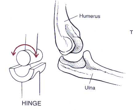
4. Hinge joints is when the convex surface of one bone fits into the concave surface of another bone, like the elbow. This joint can also be found in the phalanges and permits movement only in one plane, like a door hinge.
5. Pivot Joint is when the cylinder surface of one bone rotates within a ring formed of bone and ligament. Movement is limited to the rotation around the central axis.

6. Saddle Joint forms between bones whose articulating surfaces have both concave and convex regions. The surface of one bone fits the complementary surface of the other.

_ _ _ _ _ _ _ _ _ _ _ _ _ _ _ _ _ _ _ _ _ _ _ _ _ _ _ _ _ _ _ _ _ _ _ _ _ _ _ _ _ _ _ _ _ _ _ _ _ _ _ _ _ _ _ _ _ _ _ _ _ _ _ _ _ _ _ _ _ _ _ _ _ _ _ _ _ _ _ _ _ _ _ _ _ _ _ _ _ _
Fibrous Joints:
These are joints which connect bones but are immoveable. There are three types: sutures, syndesmosis, and gomphosis.

Structure:
Fibrous joints are made up of collagen, and they have no joint cavity.
Types of Fibrous Joints and Examples:
1. Sutures: The fibrous joints make up the sutures of the skull. Slight movement is of the joints is possible with infants because they don't lock until after birth.

2. Syndesmosis: These joints are located in between the long bones, they connect the tibia and fibula, and the radius and the ulna. Though they are slightly moveable they aren't as moveable as synovial joints.

3. Gomphosis: They also hold the teeth into the sockets of the jaw bones.

_ _ _ _ _ _ _ _ _ _ _ _ _ _ _ _ _ _ _ _ _ _ _ _ _ _ _ _ _ _ _ _ _ _ _ _ _ _ _ _ _ _ _ _ _ _ _ _ _ _ _ _ _ _ _ _ _ _ _ _ _ _ _ _ _ _ _ _ _ _ _ _ _ _ _ _ _ _ _ _ _ _ _ _ _ _ _ _
Cartilaginous joints
These joints allow limited movement when they are bent forward, to the side, or twisted. They consist of hyaline cartilage and fibrocartilage. The layers of hyaline are known as sychondrosis joints. A sychondroses joint is a rigid cartilaginous bridge between two articulating bones. These are the strongest joints in the body.

Cartilaginous joints separate the vertebrae in the vertebral column. The intervertebral discs are composed of fibrocartilage surrounding a gelatinous core. The disc absorbs shock and equalizes pressure between the adjacent vertebrae during body movement.


A cartilaginous joint is classified as an immovable joint called a synarthrosis, and a slightly movable joint a symphysis.
Types of Cartilaginous Joints and Examples:
At a synarthrosis the edges of bones are very close together to the point where they may interlock. These joints are extremely strong and are found in places where movement must be prevented, like the sutures of a skull.

At a symphysis, articulating bones are kept separate by a wedge of fibrocartilage. It is known as a amphiarthroses which allows more movement than a synathrosis but is stronger than
a freely movable joint. Every symphysis except two of them lie in the vertebral column.

_ _ _ _ _ _ _ _ _ _ _ _ _ _ _ _ _ _ _ _ _ _ _ _ _ _ _ _ _ _ _ _ _ _ _ _ _ _ _ _ _ _ _ _ _ _ _ _ _ _ _ _ _ _ _ _ _ _ _ _ _ _ _ _ _ _ _ _ _ _ _ _ _ _ _ _ _ _ _ _ _ _ _ _ _ _ _ _
Types of joint movement:
1. Flexion: When parts of a joint bend so the angle between them increases and the parts become closer to each other (ex. Bending the knee)
2. Extension: When parts of a joint straighten so they angle between them increases and the parts become further from each other (ex. Straightening the knee) 
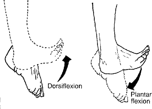
3. Dorsiflexion: When the foot bends at the ankle towards the shin
4. Plantar flexion: When the foot bends at the ankle towards the sole
5. Hyperextension: When the joint bends past its anatomical position (ex. Bending the head backwards past the upright position).jpg)
6. Abduction: Moving part of the body away from the midline (ex. Lifting the upper limb to a 90 degree angle with the side of the body)
7. Adduction: Moving part of the body towards the midline (ex. Moving the upper limb from its horizontal position back to the side of the body)

8. Rotation: Moving a joint around an axis (ex. Twisting the head from side to side)

9. Circumduction: Moving part of the body so that the end follows a circular path (ex. Moving the finger in a circle without moving the hand)

10. Pronation: Turning the hand so the palm is facing downward
11. Supination: Turning the hand so the palm is facing upward

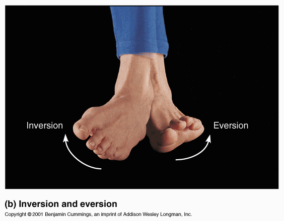
12. Eversion: Turning the foot so the sole faces laterally
13. Inversion: Turning the foot so the sole faces medially
14. Retraction: Moving a part backward (ex. Pulling the chin backwards)

15. Protraction: Moving a part forward (ex. Moving the chin forward)

16. Elevation: Raising a body part (ex. Shrugging the shoulders)

17. Depression: Lowering a body part (ex. Drooping the shoulders)
Comments (1)
Judy Hoyle said
at 2:31 pm on May 11, 2011
Great Job
You don't have permission to comment on this page.